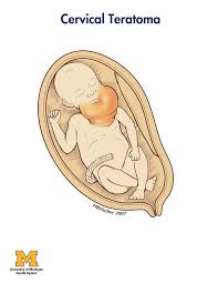
Cervical Teratoma
When a non-cancerous or benign tumor appears, it makes the baby’s neck huge and forms mass in the neck. Fetal teratoma is completely different as it matures fast and cytodifferentiation is seen half of the time, and adult teratoma is cancerous or malignant mostly. It is a very rare condition but a severe one, the tumor grows instantly and expands the size of the neck, proportion of neck along with observed polyhydramnios. The areas covered in cervical teratoma are nasopharynx and thyrocervical. Though it is attached with thyroid gland it doesn’t start forming from it. Cartilage, neural tissues and respiratory epithelium are responsible. Possibility of brain teratoma is there in some cases. Very huge tumors can lead to anemia and heart failure. Read More
Top Doctors For Cervical Teratoma Treatments


Top Hospitals For Cervical Teratoma Treatments


Cervical Teratoma
Table of contents
What is cervical teratoma?
A cervical teratoma is a rare congenital benign (non-cancerous) tumor in a baby’s neck. Cervical teratoma is a tumor that tends to be large, disfiguring masses- which are partly solid and partly fluid. The main concern of these tumors is they block the baby’s airway and food pipe (esophagus), resulting in the accumulation of fluid that can affect the baby’s growth and even result in preterm delivery.

Causes
The exact cause of cervical teratoma is idiopathic (not known). However, cervical tumors are basically germ cell tumors. Germ cells are the cells that develop into an embryo, and later on, these cells make up the reproductive systems of men and women. An old theory suggests that cervical teratomas are caused by an inability of the cells to develop into a complete body or abnormal development of a conjoint twin.
How is cervical teratoma diagnosed?
Doctors will suggest the mother undergo numerous tests to help confirm the diagnosis. Primary tests include:
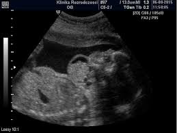
- Routine prenatal ultrasound scan- this test is usually performed around the 18th week of pregnancy. Scanning is done to rule out any sign of abnormality in the fetus.
- The diagnosis of cervical teratoma is based on thorough clinical evaluation, identification of characteristic physical findings, and detailed patient medical history and family history.
- FNA (fine needle aspiration)- to confirm the diagnosis of cervical teratoma, the doctor performs an FNA test, in which a fine needle is passed through the skin and inserted into the nodule to draw a small tissue sample. The collected tissue is then studied under a microscope.
- Biopsy- during this procedure, the doctor surgically removes a tissue sample and sends it for pathological studies.
- Other scans include CT scans (computed tomography), MRI scans(magnetic resonance imaging), and x-ray scans.
- Other laboratory tests
How is cervical teratoma treated?
The treatment option for cervical teratoma depends upon various factors such as the primary tumor’s location, stage of cancer, degree (grade) of the tumor, and whether the tumor has spread to the lymph nodes or to other organs in the body.
- In most patients, cervical teratoma is treated surgically by removing the tumor and the affected tissue. Since most teratomas are seen in children, delay in treatment can compress the windpipe (trachea), so the tumor must be removed as soon as possible.
- If these tumors are detected prenatally (during pregnancy), a procedure known as EXIT (ex-utero intrapartum treatment) can be performed. During this procedure, the baby is delivered through C-section (cesarean). The baby is attached to the placenta so that normal blood flow exchange occurs while the surgeon performs the necessary surgical procedure.
- In a few cases, the doctor might perform a tracheostomy (a surgical procedure in which an incision is made in the windpipe) to create a temporary opening to allow air passage.
- Surgical management of cervical teratoma may involve the removal of a portion or entire thyroid gland. In such cases, the patient must undergo hormone replacement therapy to obtain the hormones normally produced by the thyroid gland.
How common is Cervical Teratoma?
This is a very rare type of cervical cancer, and it is said that 80% of the cases are neonatal teratoma. The incidence is high in females compared to men. Approximately 2-3 infants in 30,000 to 40,000 live births get affected by cervical teratoma.
Symptoms
Symptoms are directly proportional to the size of the tumor. In small tumors, few symptoms are seen; in large tumors, more symptoms are seen. A few of the symptoms are listed below:
- Shortness of breath or dyspnea
- Stridor
- Trouble in swallowing
- Oesophagus (food pipe) shrinks
- Windpipe or trachea contracts
- Amniotic fluid increases
Ovarian teratoma Symptoms
This occurs only in females, and symptoms include :
- Back pain
- Frequent uterine bleeding
- Trouble in the gastrointestinal system
- Pain in Abdomen
- Swelling
FAQ
In most cases, cervical teratomas are benign. However, early diagnosis and early treatment will result in a good prognosis. The survival rate of the patients after surgical resection is >80%. The recurrence rate of cervical teratoma is rare.
Lung teratoma is uncommon, even more than cervical teratoma. However, around 300-500 cases are reported to date, and this usually happens in the left lung and happens because of the formation of abnormal germ layers.
When the disturbance in the growth hormones crosses the limit in germ cells, teratomas form.
This affects only 1-2% of people, commonly seen in infants and children. This is a subtype of extragonadal teratoma.
Brain teratoma is an extremely rare condition, and it affects 1 in 1,000,000 population. Cystic and calcified components destroy brain functioning and its growth and result in brain teratoma.
During pregnancy, a frequent ultrasound scan is recommended to monitor the condition of the fetus (baby) and amniotic fluid volume, monitor tumor size, and check the fetus’s airway system and growth. The patient is advised for a cesarean section, and the EXIT treatment option is followed in an emergency.
The most common complications of cervical teratomas are listed below:
i. The cervical teratomas can interfere with the development of the fetus growth.
ii. It can interfere with the patient’s respiration by blocking or narrowing the airway.
iii. The tumor can compress the esophagus, resulting in difficulty swallowing food or liquids.
iv. Cervical teratomas can be fatal if left untreated or if there is a delay in managing the tumor.