Osteochondrosis
Osteochondrosis is a family of self-limiting development disorders. It affects the epiphysis (the end part of the bone, which usually forms the part of the joint) and/or the apophysis (site of tendon or ligament attachment) Read More
Top Doctors For Osteochondrosis Treatments


Top Hospitals For Osteochondrosis Treatments


Osteochondrosis
In children, the skeletal system is in a constant state of development as they move into the early stages of adulthood.
The process of bone development works at the ossification centres, which are the areas where the bone grows with the help of bone-forming cells called osteoblasts. Osteochondrosis is a family of self-limiting development disorders. It affects the epiphysis (the end part of the bone, which usually forms the part of the joint) and/or the apophysis (site of tendon or ligament attachment). Osteochondrosis is seen most commonly in children and adolescents. It may, in extreme cases, also be found in adults with patients complaining about pain and disability. On radiographs, these disorders are seen as fragmentation, collapse, sclerosis, and re-ossification of osseous centres (1).
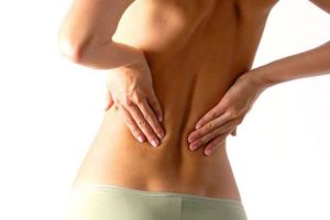
Picture Courtesy: www.mybodypain.com
Types of Osteochondrosis
Since there is a variety of diseases that can be classified as osteochondrosis, they are categorized into three main types based on the area they occur. They are articular, physeal, or non-articular. A few of the diseases under these categories have also been mentioned.
Articular diseases:
Usually affects the joint region, primarily the epiphyseal region. They include Legg-Calve-Perthes disease (affects the hip), Panner’s disease (affects the elbow), Frieberg’s disease (affects the second toe), and Kohler disease (affects the foot).
- Legg-Calve-Perthes disease: Occurs mostly in children between the ages of 4 and 8 with the chances of boys getting affected being more than girls [10]. Incidence of the disease is high in patients with low birth weight, hereditary, abnormal birth presentation, lower socioeconomic status (due to lack of nutritious food intake) [11]. This disease affects the hip region, where the thighbone (femur) and pelvis meet in a ball and socket joint. This disease is said to take place when blood is temporarily cut off to the femoral head which forms the ball part to the socket. Thus, leads to the slow death of the bone, meaning that it breaks more easily and heals poorly (2). The most common presentations are hip deformity, referred pain in the knee, limping. On physical examination, limited hip abduction (movement of the hip away from the midline of the body), the difference in the leg length, and restriction of internal rotation will be observed. Radiographs generally show a varying degree of fragmentation, flattening, sclerosis with joint space widening of the proximal femur growth centre. The risk of developing arthritis is high at a young age if not treated early.
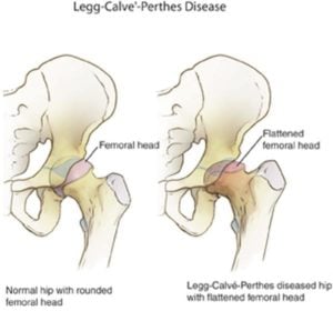
Picture Courtesy: www.epharmapedia.com
- Panner’s Disease: Affects the growth plate of children aged less than ten. The growth plate defines the final size of the bone. Blood flowing to these plates is restricted, causing the plates to die. Most often affects the dominant arm and is triggered by some trauma caused to the elbow (3). Patients are usually presented with pain in the lateral side of the elbow. Radiographic image shows fragmentation and slit in the humeral capitellum. In most cases, it can be healed with rest and conservative management (like pain killers).
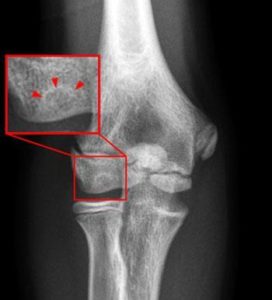
Picture Courtesy: www.radsource.us
- Frieberg’s Disease: Commonly seen in young females around the age of eighteen and affects the metatarsal bones (2nd most common followed by 3rd and 4th) [12] in the foot. It is an extremely rare condition (4). Patients are presented with severe pain associated with swelling and tenderness of the affected metatarsal bone. Radiographic image shows a varying degree of flattening and abnormal hardening of the affected articular surface [13]
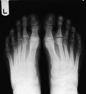
Picture Courtesy: www.nwhealth.edu
- Kohler disease: Affects the navicular bone of the foot. It usually affects children between the ages of three and five, mostly young boys rather than girls5). Patients are generally presented with pain in the midfoot associated with swelling and warmth. During the physical examination, tenderness will be present on the navicular bone [14]. Radiographic images show fragmentation, flattening, and sclerosis of the navicular bone [15].
Physeal diseases:
It is a type of osteochondrosis that affects the vertebral joints of growing children (Scheuermann’s Kyphosis) and the long bones (Blount disease).
- Scheuermann’s Kyphosis: AKA Juvenile Kyphosis, is a condition where the vertebral bones grow at a rate different from each other, eventually giving the back a hump shape look. Usually seen in children who just enter puberty, this disorder affects either the thoracic spine (upper back) or the lumbar spine (lower back). A Scheuermann’s kyphosis is diagnosed when:
There is a curvature of forty-five degrees or more.
Three or more vertebrae wedged together by at least 5 degrees per segment.
Symptoms usually include pain during physical activities, muscle cramps, spasms, tiredness, muscle stiffness, et cetera (8).
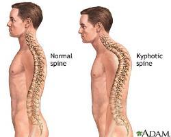
Picture Courtesy: www.s-media-cache-ak0.pinimg.com
- Blount Disease: Involves the shin bone (bone below the knee) bending inwards and looking like a bow leg. Usually seen in children and adolescents, there are also cases where adults have been affected by it. It may cause intense pain in teenagers. It is more prevalent in African-American children and has also been related to obesity (9).
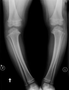
Picture Courtesy: www.images.radiopaedia.org
Non-articular diseases:
The diseases that fall under this category can affect any part of the skeleton, mostly in regions of tendon and ligament attachment. An example is Sever’s disease (AKA calcaneal apophysitis) which affects the heels (6). Sever’s Disease is a common heel injury occurring in kids. Though painful, it has no long-term effects and is temporary. It is caused due to the swelling of the growth plate of the heel (7).
Symptoms and Treatments
The most common symptom of Osteochondrosis is a pain in the affected region either due to physical activity or if pressure is applied. Other symptoms include tenderness, swelling, joint dysfunction, and/or an inability to fully straighten the affected limb.
Osteochondrosis, on the bright side, can be easily diagnosed as it shows up clearly on x-ray tests. Note-worthy is that no clear-cut cure currently exists to stop and reverse the damage caused by osteochondrosis. So, the goal of any treatment is to slow down the damage process and control and ease the symptoms associated with the disease. This would involve educating the patients about the disease and the measures to take to avoid any further damage (avoiding contact sports etc.).
Braces might also be recommended along with exercise and physiotherapy. In certain cases, when conservative methods are ineffective, doctors may recommend surgery when they feel that such procedures can help the patient recover better and in a guided manner, though this is usually in extreme cases. The surgeries and treatment provided are on a case basis. Some of the surgeries performed are:
Arthroscopic procedures are minimally invasive surgeries performed on the joint.
Shelf acetabuloplasty procedures to provide lateral support.
And in extreme cases, salvage and reconstructive surgeries are done to reduce disability.
Works Cited
1. Varshney, Manish Kumar. Osteochondroses. Medscape.com. [Online] [Cited: May 02, 2017.] http://emedicine.medscape.com/article/1254668-overview#a1.
2. Mayo Clinic Staff. Legg-Calve-Perthes disease. MayoClinic.org. [Online] [Cited: May 02, 2017.] http://www.mayoclinic.org/diseases-conditions/legg-calve-perthes-disease/basics/definition/con-20035572.
3. Kids Health. Panner’s Disease. Kidshealth.org. [Online] [Cited: May 02, 2017.] http://kidshealth.org/en/parents/panners-disease.html?WT.ac=ctg.
4. Imm, Dr Nick. Freiberg’s Disease. Patient.info. [Online] [Cited: May 02, 2017.] https://patient.info/in/doctor/freibergs-disease.
5. Weaver, Dr. Benjamin. Kohler’s Disease — Symptoms, Treatment, and Prognosis. FootVitals.com. [Online] [Cited: May 02, 2017.] http://www.footvitals.com/bones/kohlers-disease.html.
6. Bowman, Joe. What Are Osteochondroses? healthline.com. [Online] [Cited: May 02, 2017.] http://www.healthline.com/health/osteochondroses#types2.
7. Kids Health. Sever’s Disease. KidsHealth.org. [Online] [Cited: May 02, 2017.] http://kidshealth.org/en/parents/severs-disease.html.
8. McAfee, Paul C. Scheuermann’s Disease of the Thoracic and Lumbar Spine. spine-health.com. [Online] [Cited: May 27, 2017.] https://www.spine-health.com/conditions/spinal-deformities/scheuermanns-disease-thoracic-and-lumbar-spine
9. KidsHealth.org. Blount DIsease. kidshealth.org. [Online] [Cited: May 27,2017.] http://kidshealth.org/en/teens/blount-disease.html#
10. Barker DJ, Hall AJ. The epidemiology of Perthes’ disease. Clin Orthop Relat Res. 1986;(209):89–94. Nigrovic PA. Overview of hip pain in childhood. Online.http://www.uptodate.com/online/content/topic.do?topicKey=gen_pedi/21233 Accessed October 25, 2010.
11. Hall AJ, Barker DJ. Perthes’ disease in Yorkshire. J Bone Joint Surg Br. 1989;71(2):229–233. Kenet G, Ezra E, Wientroub S, et al. Perthes’ disease and the search for genetic associations: collagen mutations, Gaucher’s disease, and thrombophilia. J Bone Joint Surg Br. 2008;90(11):1507–1511.
12. Katcherian DA. Treatment of Freiberg’s disease. Orthop Clin North Am. 1994;25(1):69–81.
13. Kasser JR. The foot. In: Lovell WW, Winter RB, Morrissy RT, Weinstein SL, eds. Lovell and Winter’s Pediatric Orthopaedics. 6th ed. Philadelphia, Pa.: Lippincott Williams & Wilkins; 2006:1257–1328.
14.. Tsirikos AI, Riddle EC, Kruse R. Bilateral Köhler’s disease in identical twins. Clin Orthop Relat Res. 2003;(409):195–198. Borges JL, Guille JT, Bowen JR. Köhler’s bone disease of the tarsal navicular. J Pediatr Orthop. 1995;15(5):596–598.
15. Borges JL, Guille JT, Bowen JR. Köhler’s bone disease of the tarsal navicular. J Pediatr Orthop. 1995;15(5):596–598.

FAQ
- What causes bones to enlarge?
The osteoblasts (one of the bone cells responsible for bone formation and remodeling) become excited and begin to proliferate at abnormal uncontrolled levels. This causes the enlargement of the quickly formed bone. These abnormally proliferating bone cells tend to have more blood circulation than normal cells. The cause of the excitation of the osteoblasts is not known.